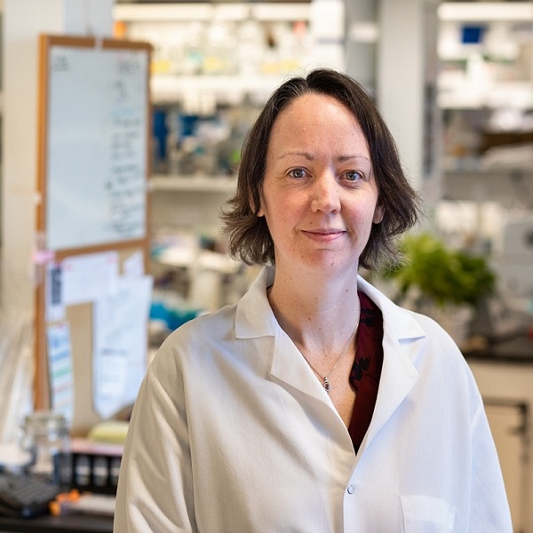
Celeste Nelson explores how branching patterns emerge during development. Her research combines biology with engineering and computational modeling — with the ultimate goal of building functional tissues outside the body.
“These branched architectures are ubiquitous,” said Nelson, a professor of chemical and biological engineering. “In the lung, they’re important for making sure there’s enough surface area to move oxygen from a breath into the bloodstream,” while in organs like the mammary or salivary glands, “it’s kind of a space-filling mission: making enough milk, making enough saliva.”
Much of Nelson’s work is focused on the forces that cause a clump of cells in the embryo to grow into the tree-like pattern of airways in the newborn lung. To understand how different physical processes can achieve similar results, her group has investigated lung development not only in mice, a model for human development, but also in birds and reptiles.
Nelson’s lab has spent years uncovering the details of mouse lung development, finding that physical signals from smooth muscle cells direct cells lining the developing airway to split into two branches — a process that occurs millions of times to form each well-functioning newborn lung. And more recent work led by graduate student Katie Goodwin has shown that smooth muscle cells also shape growth of the very earliest airway branches, which form when a bud begins to grow on the side of a main branch (versus the end of a branch splitting in two).
Reptile lungs, in contrast, are simpler sacs that lack elaborate branching patterns. In developing embryos, smooth muscle cells form a hexagonal mesh that wraps around the growing lung, contracting and squeezing the epithelial cells to form the folds of the mature organ. In a collaboration with Andrej Košmrlj, an assistant professor of mechanical and aerospace engineering, and Jared Toettcher, an assistant professor of molecular biology, Nelson has received support from Princeton’s Eric and Wendy Schmidt Transformative Technology Fund to develop a 3-D printing technology for artificial organs based on knowledge of reptile lung development.
“Can we take what we’ve learned from looking beyond conventional model systems — learning how various species build their lungs — to come up with new ways to engineer three-dimensional structures outside of the body?” Nelson asked. “We’ll see how far we can go with this idea of one contractile tissue like muscle pushing on another to force it to change shape.”
Source: engineering.princeton


